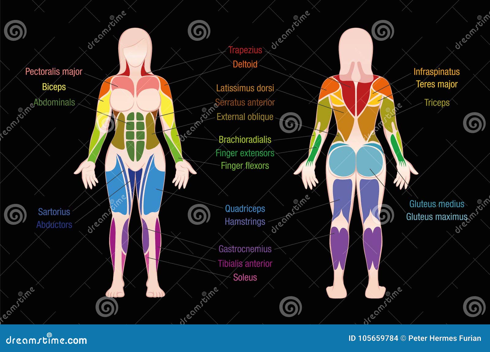
Leg Muscles Diagram Labeled : Anatomical Model Labeled Ppt Download / This diagram depicts anatomy of leg muscles with parts and labels.. Medial compartment, also known as adductor compartment; Our goal is that these leg anatomy worksheets pictures gallery can be a direction for you, bring you more references and also make you have a great day. This is an online quiz called leg muscle labeling. This image is titled nerves of the leg diagram and is attached to our article about leg nerves and reflex motion in feet. Leg muscles labeled take a look at the leg muscles diagram below, where you see each muscle clearly labeled.
You may also find vastus intermedius deep layer, vastus medial. The thigh (proximal lower limb) muscles are arranged into three compartments : This diagram depicts power leg muscle group.human anatomy diagrams show internal organs, cells, systems, conditions, symptoms and sickness information and/or tips for healthy living. The gastrocnemius muscle has two large bellies, called the medial head and the lateral head, and inserts into the calcaneus bone of the. Your legs are two of your most important body parts.
Muscle Names Stock Illustrations 50 Muscle Names Stock Illustrations Vectors Clipart Dreamstime from thumbs.dreamstime.com They're responsible, respectively, for extending your foot (pointing your toes) and flexing your foot (pulling your foot towards your shin). Part of the teachme series. The aim of this exercise is to improve your confidence in identifying different structures. It helps maintain erect posture, abducts the thigh, and rotates the thigh outward. The 3 muscles are called triceps coxae. This image is titled nerves of the leg diagram and is attached to our article about leg nerves and reflex motion in feet. The hamstring muscles, also known as the rear thighs, make up the backside of the upper leg anatomy. Biceps femoris (long head) biceps femoris (short head) semitendinosus.
Muscle anatomy head 12 photos of the muscle anatomy head dog head muscle anatomy, human.
The thigh (proximal lower limb) muscles are arranged into three compartments : Add to playlist 24 playlists. It dorsi flexes and inverts the foot, supports the arch, and is controlled by the deep peroneal. Medial compartment, also known as adductor compartment; Posted on may 19, 2016 by admin. Anterior and posterior muscles of upper leg. Gastrocnemius muscle, large posterior muscle of the calf of the leg. Anterior compartment, also known as the extensor compartment; It originates at the back of the femur (thighbone) and patella (kneecap). Muscles in the posterior compartment of the leg. The muscles that make up the quadriceps are the strongest and leanest of all muscles in the body. Your legs are two of your most important body parts. This diagram depicts anatomy of leg muscles with parts and labels.
As these muscles contract and relax, they move skeletal bones to create movement of the body. It helps maintain erect posture, abducts the thigh, and rotates the thigh outward. Anterior and posterior muscles of upper leg. Your legs are two of your most important body parts. Home › training design › anatomy and physiology › muscle charts of the human body.
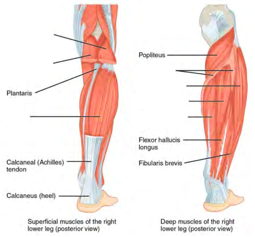
Free Anatomy Quiz Muscles Of The Lower Limb Anterior Locations Quiz 1 from www.free-anatomy-quiz.com You need to get 100% to score the 10 points available. The legs include the upper leg, knee, lower leg, ankle, and. Anterior and posterior muscles of upper leg. In this image, you will find muscle of the human leg diagram, hip and femur middle layer, hip and femur deep layer, overview of the most important muscles of the leg, femur middle layer, femur deep layer, rectus femoris m. The hamstring muscle attachment points. Muscles in the posterior compartment of the leg. It acts as a tensor of the arches of the foot, but can also be added with the first digit and plantar flexion of its first phalanx. As far as the lower leg muscle anatomy goes, the major muscles include two calf muscles and one shin muscle.
We'll break down the anatomy and function of the upper leg, knee, lower leg.
The hamstring muscle attachment points. They allow you to move and provide support for your upper body. Leg muscles anatomy leg anatomy human body anatomy human anatomy and physiology muscle anatomy anatomy study anatomy models muscular system medical anatomy. An extensor muscle that straightens or lifts the foot. Your legs are two of your most important body parts. It acts as a tensor of the arches of the foot, but can also be added with the first digit and plantar flexion of its first phalanx. Home › training design › anatomy and physiology › muscle charts of the human body. The medical information on this site is provided as an information resource only, and is not to be used or relied on for any. This is an online quiz called leg muscle labeling. Below the gluteus maximus is the smaller gluteus medius. One of the most important tendons in terms of mobility of the leg is the achilles tendon. Gastrocnemius muscle, large posterior muscle of the calf of the leg. Learn vocabulary, terms, and more with flashcards, games, and other study tools.
Posted on may 19, 2016 by admin. For women, shaping the thigh muscles is an essential goal of physical fitness. An extensor muscle that straightens or lifts the foot. You may also find vastus intermedius deep layer, vastus medial. It dorsi flexes and inverts the foot, supports the arch, and is controlled by the deep peroneal.
Pre Lab 10 Human Anatomy Lab Manual from uta.pressbooks.pub Muscles and articulations of the human body. This diagram depicts power leg muscle group.human anatomy diagrams show internal organs, cells, systems, conditions, symptoms and sickness information and/or tips for healthy living. This image is titled nerves of the leg diagram and is attached to our article about leg nerves and reflex motion in feet. It helps maintain erect posture, abducts the thigh, and rotates the thigh outward. The largest of them is the most superficial muscle, the gluteus maximus. The muscles of the lower leg, called simply the leg by anatomists, largely move the foot and toes. You may also find vastus intermedius deep layer, vastus medial. Muscle anatomy head 12 photos of the muscle anatomy head dog head muscle anatomy, human.
Muscle anatomy head 12 photos of the muscle anatomy head dog head muscle anatomy, human.
The 3 muscles are called triceps coxae. One of the most important tendons in terms of mobility of the leg is the achilles tendon. Posted on may 19, 2016 by admin. Related posts of lower leg muscles diagram muscle and bone anatomy. Part of the teachme series. The majority of muscles in the leg are considered long muscles, in that they stretch great distances. Muscle and bone anatomy 12 photos of the muscle and bone anatomy back muscles and bones anatomy, human muscle and bone anatomy, muscle & bone anatomy 3d free download, muscle and bone anatomy app, muscle and bone anatomy quiz, human muscles, back muscles and bones anatomy, human muscle and bone anatomy, muscle & bone. Observe the leg muscle diagram posted above and notice that there are many parts in the muscles.the largest muscle masses in the leg are present in the thigh and the calf. An extensor muscle that straightens or lifts the foot. The muscles of the lower leg, called simply the leg by anatomists, largely move the foot and toes. By the way, related with muscle labeling worksheet, we already collected particular variation of pictures to inform you more. Below the gluteus maximus is the smaller gluteus medius. The following diagram illustrates the actions of the terms adduction, abduction, flexion and extension at the different joints.
The following diagram illustrates the actions of the terms adduction, abduction, flexion and extension at the different joints leg muscles diagram. Add to playlist 24 playlists.


0 Komentar